Diagnostic Imaging Glossary
Glossary of Terms for Diagnostic Imaging
This glossary is made to learn new terms about my futur job, which is diagnostic imaging.
- Magnetic Resonance Imaging (MRI)
- noun
- Magnetic resonance imaging (MRI) is a medical imaging technique that uses a magnetic field and computer-generated radio waves to create detailed images of the organs and tissues in your body.
- Example: However CA15-3 is still relatively non-specific as elevated levels may be found in bronchial, ovarian and colorectal tumours, and non-malignant conditions such as cirrhosis, hepatitis and benign breast disease (38). 5.5 Future Investigations The use of magnetic resonance imaging (MRI) in breast cancer is being examined in several distinct settings.
- fr: Imagerie par résonance magnétique (IRM)

- Computed Tomography
- noun
- The term “computed tomography”, or CT, refers to a computerized x-ray imaging procedure in which a narrow beam of x-rays is aimed at a patient and quickly rotated around the body, producing signals that are processed by the machine's computer to generate cross-sectional images—or “slices”—of the body.
- Example: Future directions for detection of coronary reperfusion after thrombolytic therapy Coronary echocardiography, Magnetic resonance imaging (MRI), Cardiac computed tomography scanner (CT) and transthoracic Doppler echocardiography (TTDE) are just some of the emerging techniques used by clinicians to accurately evaluate IRA patency, providing information on coronary reperfusion.
- fr: Tomodensitométrie

- Barium enema
- noun
- A procedure in which a liquid that contains barium sulfate is put through the anus into the rectum and colon. Barium sulfate is a silver-white metallic compound that helps show pictures of the colon, rectum, and anus on an x-ray.
- Example: Double contrast barium enema : Usually reveals the growth as either a stricture or a filling defect (apple-core deformity). However, it is important to remember that a negative barium enema doesn't definitely exclude the presence of a small tumour, particularly in the presence of extensive diverticulosis.
- fr: Lavement baryté

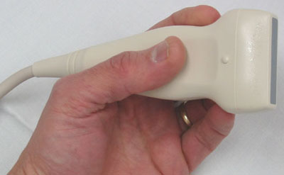
- Ultrasound
- noun
- An ultrasound scan is a medical test that uses high-frequency sound waves to capture live images from the inside of your body.
- Example: A mass in Mary's appendix was detected and investigated using ultrasound and a CT scan, she was observed for 3 days and treated with intravenous antibiotics which reduced the mass, surgery was not thought necessary so Mary was discharged and a follow-up outpatient appointment was arranged in a month's time.
- fr: Échographie
- Abdominal aortic aneurysm
- noun
- An abdominal aortic aneurysm is an enlarged area in the lower part of the major vessel that supplies blood to the body (aorta). The aorta runs from your heart through the center of your chest and abdomen.
- Example: Although it is impossible to determine the precise cause of the emboli without further investigation, there was no evidence to suggest the presence of atrial fibrillation or an abdominal aortic aneurysm.
- fr: Anévrisme de l'aorte abdominale

- Diverticula
- noun
- Diverticula are small, bulging pouches that can form in the lining of your digestive system. They are found most often in the lower part of the large intestine (colon).
- Example: However, it is important to remember that a negative barium enema doesn't definitely exclude the presence of a small tumour, particularly in the presence of extensive diverticulosis.
- fr: Diverticules
- Urography
- noun
- Urography is an examination used to evaluate the kidneys, ureters and bladder. Excretory urography, also known as intravenous pyelogram, is performed using conventional x-ray after the intravenous administration of radiographic contrast material.
- Example: Tumours, tuberculous abscesses, kidney stones, and obstruction by prostatic enlargement may be detected by this method. Specific types of urography include pyelography (examination of the kidney and ureter) and cystography (examination of the bladder).
- fr: Urographie

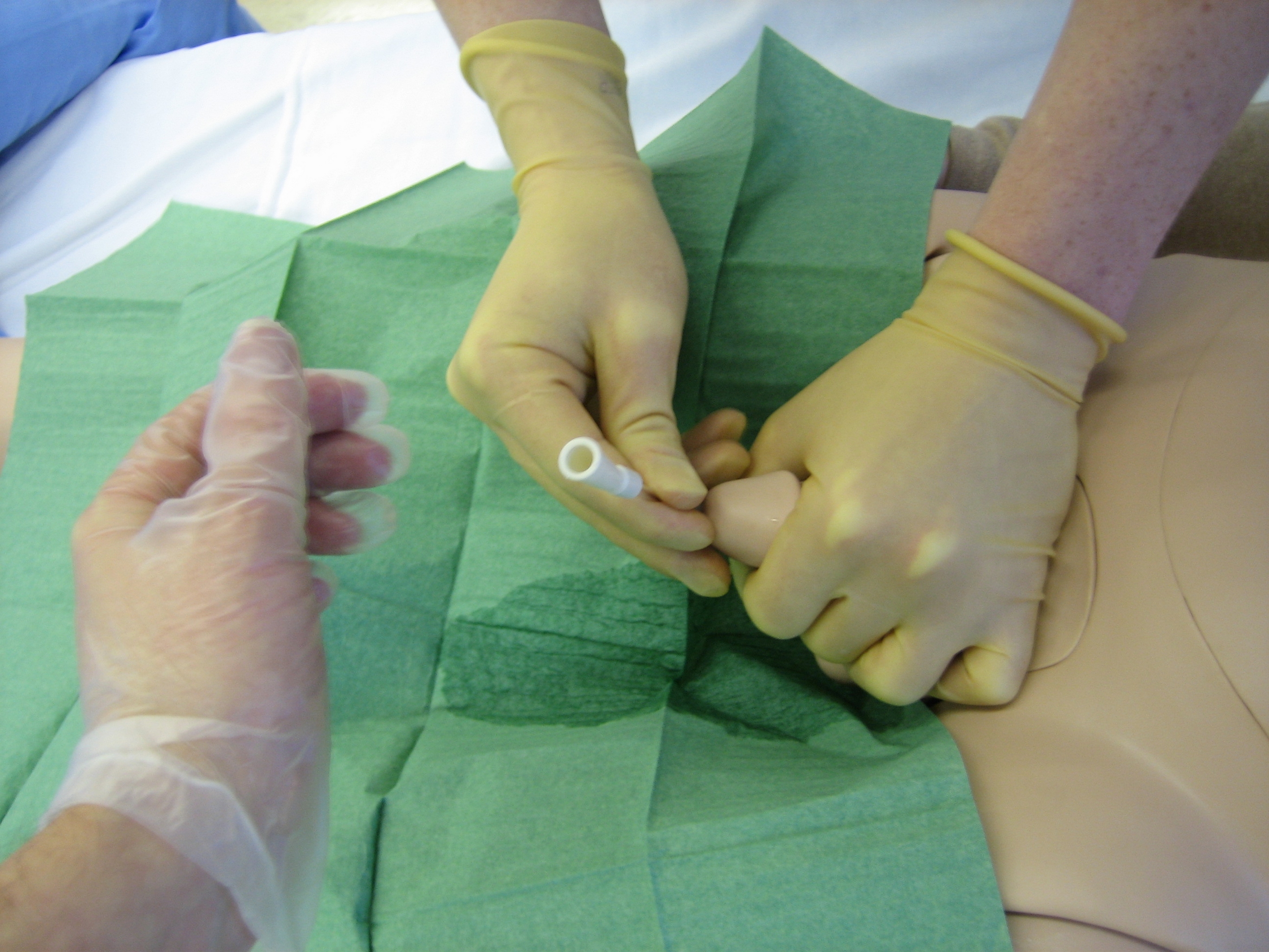
- Urinary catheter
- noun
- A urinary catheter is a tube placed in the body to drain and collect urine from the bladder.
- Example: Insert a urinary catheter to monitor resuscitation if the patient is dehydrated.
- fr: Sonde urinaire
- Appendicitis
- noun
- Appendicitis is an inflammation of the appendix, a finger-shaped pouch that projects from your colon on the lower right side of your abdomen. Appendicitis causes pain in your lower right abdomen.
- Example: Analysis of history Differentials: Acute appendicitis: The pain is describing could be appendicitis. Although in appendicitis the pain normally starts around the umbilicus and moves to the right iliac fossa (RIF), but in the pain started in the RIF.
- fr: Appendicite

- Hepatocarcinoma
- noun
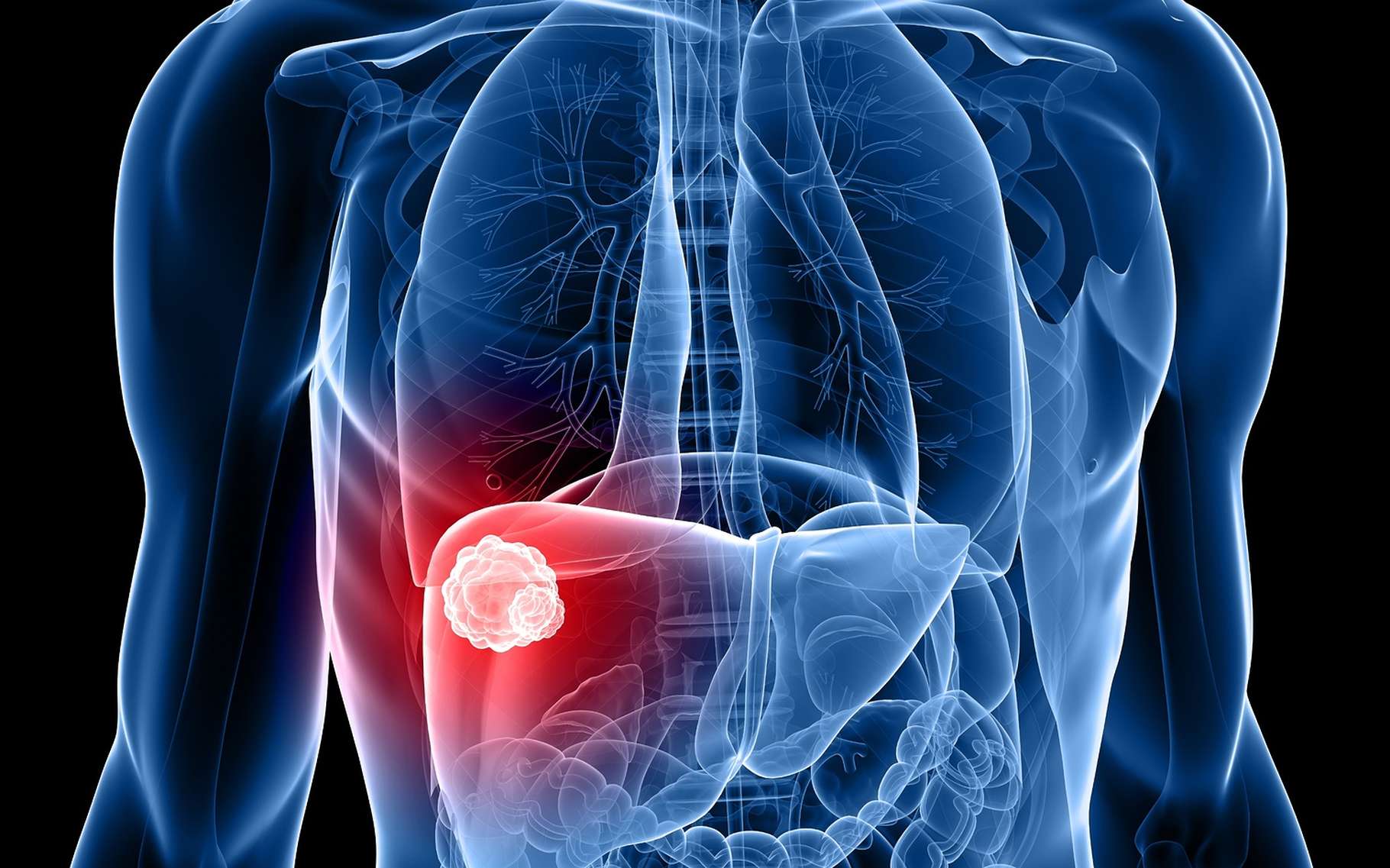
- Hepatocellular carcinoma (HCC) is the most common type of primary liver cancer. Hepatocellular carcinoma occurs most often in people with chronic liver diseases, such as cirrhosis caused by hepatitis B or hepatitis C infection.
- Example: This harmful substance, which is made by certain types of mold on peanuts, corn, and other nuts and grains, can cause hepatocellular carcinoma. The U.S. has safety measures that limit aflatoxin in the food supply.
- fr: Hépatocarcinome
- Contrast agent
- noun
- A contrast agent (or contrast medium) is a substance used to increase the contrast of structures or fluids within the body in medical imaging. Contrast agents are commonly used to improve the visibility of blood vessels and the gastrointestinal tract.
- Example: Acetylcysteine may be given as premedication for patients at risk of developing nephrotoxicity, but its efficacy is uncertain. Oral antihyperglycemic drugs, such as metformin, should be withheld for 48 hours after IV contrast administration to avoid drug accumulation if contrast-induced nephrotoxicity occurs.
- fr: Agent de contraste
- Radiology
- noun
- The science dealing with X-rays and other high-energy radiation, especially the use of such radiation for the diagnosis and treatment of disease.
- Example: Although only a proportion of these have symptoms. (2) radiology shows loss of joint space, subchondral sclerosis and cysts, marginal osteophytes.
- fr: Radiologie

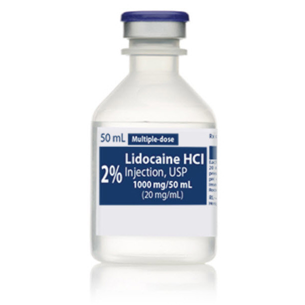
- Lidocaine
- noun
- Lidocaine is a crystalline compound C14H22N2O that is used in the form of its hydrochloride as a local anesthetic and as an antiarrhythmic agent.
- Example: Injecting your patient with hydrogen peroxide instead of lidocaine.
- fr: Lidocaïne
- X ray
- noun
- X-rays are a type of radiation called electromagnetic waves. X-ray imaging creates pictures of the inside of your body. The images show the parts of your body in different shades of black and white. The most familiar use of x-rays is checking for fractures (broken bones), but x-rays are also used in other ways.
- Example: Imaging- USS (distinguish between solid cystic swellings), CT (useful for almost any mass, especially carotid body tumours) Floor of mouth x ray (Submandibular duct calculi), Sialography (contrast study to determine possible cause and site of lesion).
- fr: Rayon X

- Hemothorax
- noun
- Hemothorax is a collection of blood in the space between the chest wall and the lung (the pleural cavity).
- Example: The symptoms of a hemothorax may include chest pain and difficulty breathing, while the clinical signs may include reduced breath sounds on the affected side and a rapid heart rate.
- fr: Hémothorax